CUSTOMER SUCCESS STORY
Two-Photon Metabolic FLIM with the Coherent Axon 780 Fibre Laser
Becker & Hickl GmbH – a technology leader in equipment for photon counting - has shown earlier that small femtosecond fibre lasers can be used as inexpensive excitation sources for multiphoton fluorescence imaging systems. With an emission wavelength of 780 nm, 40 MHz to 80 MHz pulse rate, and an average power of 100 mW to 500 mW the lasers are not only suitable for excitation of NAD(P)H but also for a wide variety of other fluorophores [1, 2], including those of extremely short fluorescence lifetime [3, 4, 5].
Becker & Hickl were therefore interested to see how the Axon femtosecond fibre laser would perform in these applications.
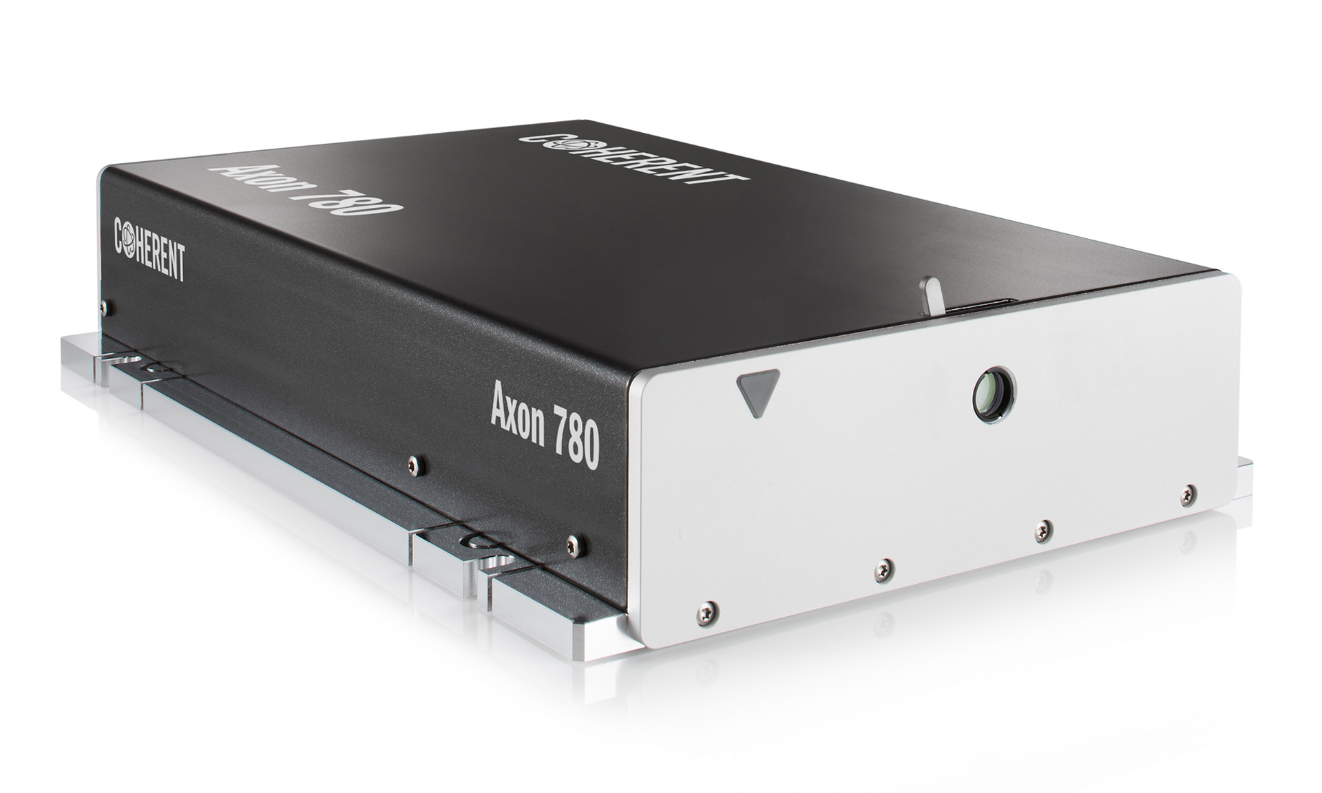
Fig. 1: Coherent Axon 780 femtosecond laser
System Architecture
As a test system they used a bh DCS-120 MP multiphoton FLIM system. The architecture of this system is shown in Fig. 2.
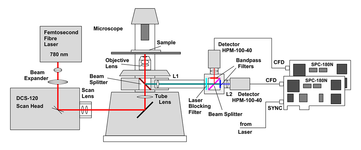
For maximum spatial resolution it is important that the laser beam completely fills the back aperture of the microscope lens. With the usual focal lengths of the scan and tube lenses this is not automatically the case. The beam is therefore expanded by a factor 1.5 before it enters the scanner. The beam diameter in the back aperture is approximately 12 mm, which is enough to fill the aperture of even the largest microscope lenses. Over-filling the aperture is unproblematic. The associated loss in excitation power can be tolerated because the laser delivers far more power than needed.
Fluorescence light from the sample is collected back through the microscope lens, and fed through a non-descanned beam path. L1 and L2 form a periscope. The periscope also collects photons which are not perfectly collimated by the microscope lens, e.g. photons which are scattered on the way out of a thick sample. The fluorescence light is split into two wavelength intervals, and detected by two bh HPM-100-40 hybrid detectors [6, 7]. The single-photon pulses from the detectors are recorded in two SPC-180N TCSPC / FLIM modules [1]. The SPC-180N modules determine the detection times of the photons after the excitation pulses and the position of the scanner in the moment of the photon detection. This information is used to build up the FLIM images. These are arrays of pixels, with each pixel containing a full fluorescence decay curve in a large number of time channels [1].
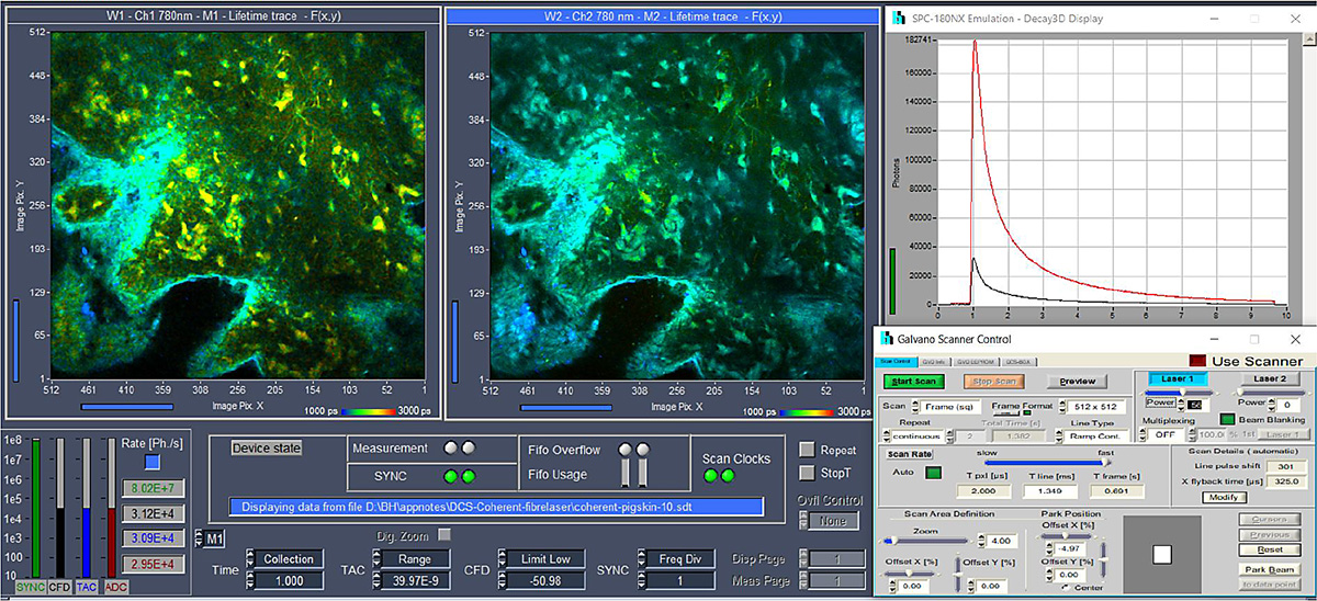
Scanning of the laser beam and beam blanking in the flyback phases are hardware-controlled by a bh GVD-140 scan controller card. Laser-intensity control is performed via the AOM control signal input of the Axon laser. This signal is provided by the GVD-140 card as well. The entire system is operated by bh's SPCM data-acquisition and control software [1], providing a fully integrated FLIM system with scanner control, laser control, data acquisition, and data analysis. The user interface of the FLIM system is shown in Fig. 3.
Result
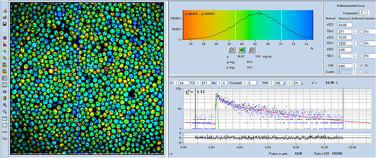
FLIM images taken with the DCS-120-AXON combination are shown in Fig. 4 through Fig. 6. An 40x, NA= 1.3 oil immersion lens was used for all images. Data analysis was performed with the bh SPCImage NG FLIM data analysis suite. Fig. 4 and Fig. 5 show colour-coded FLIM images of the mean (amplitude-weighted) lifetime and of the metabolic indicator, a1. A histogram of the selected image parameter (tm or a1) is shown upper right, a decay curve at the cursor position is shown lower right. Decay parameters at the selected spot are shown far right.

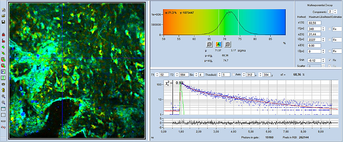
As can be seen, a1 is clearly different in different cells. This indicates that the metabolic state is different in different cells. High a1 (shown blue) indicates that the metabolism is more glycolytic, low a1 (yellow) shows that it is more oxydative.
References
- W. Becker, The bh TCSPC Handbook. 10th edition. Becker & Hickl GmbH (2023), available on www.becker-hickl.com, printed copies available from bh
- W. Becker, C. Junghans, H. Netz, Two-Photon FLIM with a Femtosecond Fibre Laser. Application note, available on www.becker-hickl.com
- W. Becker, C. Junghans, A. Bergmann, Two-Photon FLIM of Mushroom Spores Reveals Ultra-Fast Decay Component. Application note (2021), available on www.becker-hickl.com
- W. Becker, A. Bergmann, C. Junghans, Ultra-Fast Fluorescence Decay in Natural Carotenoids. Application note, www. becker-hickl.com (2022)
- W. Becker, C. Junghans, V. Shcheslavskiy, High-Resolution Multiphoton FLIM Reveals Ultra-Fast Fluorescence Decay in Human Hair. Application note, www. becker-hickl.com (2023)
- W. Becker, B. Su, K. Weisshart, O. Holub, FLIM and FCS Detection in Laser-Scanning Microscopes: Increased Efficiency by GaAsP Hybrid Detectors. Micr. Res. Tech. 74, 804-811 (2011)
- Becker & Hickl GmbH, Sub-20ps IRF Width from Hybrid Detectors and MCP-PMTs. Application note, available on www.becker-hickl.com
“We have demonstrated that the Coherent Axon 780 femtosecond fibre laser, combined with bh's precision scanner optics, detectors, TCSPC / FLIM electronics, and data analysis software delivers accurate information on the metabolism of live cells and tissues."
– Wolfgang Becker, Managing Director, Becker & Hickl GmbH, Berlin, Germany

